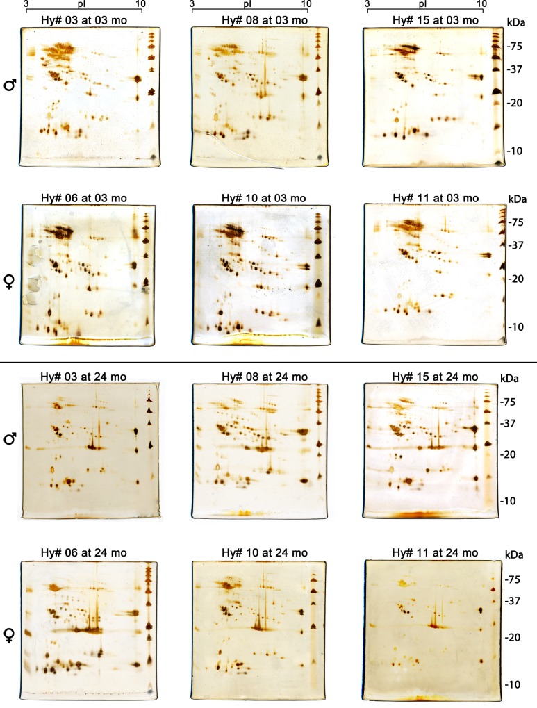
Fig 6. Comparative distribution of protein spots in two-dimensional gel electrophoresis of individual male (Hy# 03, Hy# 08 and Hy# 15) and female (Hy# 06, Hy# 10 and Hy# 11) hybrids at 3 and 24 months old (mo).
The images of the biological mother (B. erythromelas) and father (B. neuwiedi) pooled venoms are depicted in Fig 7. Gels were run under identical conditions and silver stained. For comparison, boxes were drawn over images of Hy# 03 to facilitate comparison: green dashed boxes: acidic proteins (molecular mass: 50–125 kDa, pI 4.0–6.0); blue dashed box: neutral proteins (molecular mass: 23–25 kDa, pI 6.5–7.0).

