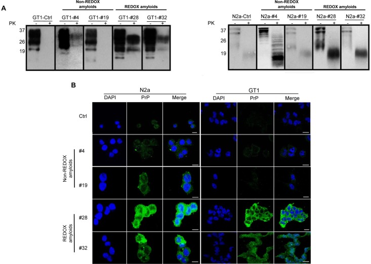
Fig 5. Seeding assay of N2a and GT1 cell cultures with synthetic amyloid fibrils.
Seeding of recMoPrP(23–231) amyloid preparations induced the conversion of endogenous PrPC to mildly PK resistant forms (A) and accumulation (B) in mouse neuroblastoma N2a and mouse hypothalamic GT1 amyloid-infected cell lines analyzed six passages after the infection (P6). Western blotting shows the partial protease K (PK) resistance of N2a and GT1 amyloid fibril-infected cell lysates. Fibril-infected cell lysates (PK-lanes) were digested with PK at ratio 1:500 (w/w) (PK+ lanes). Western blots were performed using Fab D18 monoclonal antibody (1μg/mL) for GT1 infected cells and Clone-P (1μg/mL) for N2a infected cells. Blots were developed with the enhanced chemiluminescent system (ECL, Amersham Biosciences) and visualized on Hyperfilm (Amersham Biosciences) (A). Immunofluorescence imaging shows the accumulations of PrP in N2a and GT1 amyloid fibril-infected cell lines. The deposition and level of PrP (green) in amyloid fibril-infected cell lines after six passages were detected by Fab D18 monoclonal antibody (10 μg/mL final concentration). The nuclei (blue) were stained with DAPI. Scale bar is 20μm (B).

