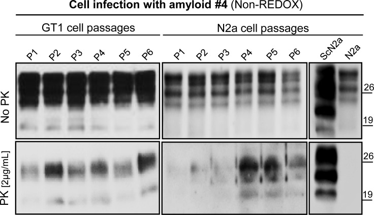
Fig 6. PK digestion assay of GT1 and N2a cells collected at different passages after infection with amyloid fibrils.
Western blotting of GT1 and N2a cell lines infected with PrP amyloid #4 was observed throughout, from first passage (P1) to sixth passage (P6) and after treatment with proteinase K at ratio 1:500 (w/w). Western blot was performed using Fab D18 monoclonal antibody (1μg/mL). Blots were developed with the enhanced chemiluminescent system (ECL, Amersham Biosciences) and visualized on Hyperfilm (Amersham Biosciences).

