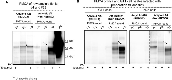
Fig 7. PMCA analysis of raw fibrils and cell lysates from infected N2a and GT1 cell lines.
Seeding ability of amyloid #4 and #28 by means of PMCA using brain homogenates of CD1 mice as substrates for amplification (A). PMCA analysis of GT1 and N2a cell lysates (infected with preparations #4 and #28) and collected at passage six (P6) after the infection (B). Black arrow indicates PK-resistant PrP. Asterisk indicates unspecific binding. Western blots were performed using 6D11 monoclonal antibody to PrP (0.2 μg/mL, Covance). Blots were developed with the enhanced chemiluminescent system (ECL, Amersham Biosciences) and visualized using a G:BOX Chemi Syngene System.

