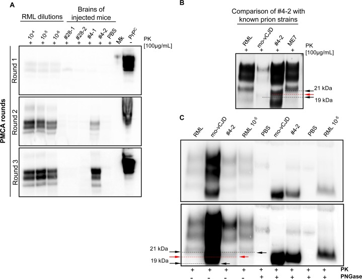
Fig 8. Amplification and characterization of a new prion isolate obtained from the brain of amyloid-infected animals.
PMCA assessment using the brain homogenate of injected animals (with amyloid #4 or #28) as seed and the brain of wild type CD1 animals as substrate. Serial dilution of RML prion strain were used as internal control for PMCA efficiency (A). Western blot (B) and PNGase comparison (C) of amplified clone #4 (PMCA-#4) with known prion strains (RML, mouse adapted vCJD and ME7). Read arrows in B and C indicate the different electrophoretic mobility of the unglycosylated PrP band of PMCA-#4 (migrating at around 20kDa) compared to that of known prion strains migrating at 19 kDa or 21 kDa (black arrows). Western blots were performed using 6D11 monoclonal antibody to PrP (0.2 μg/mL, Covance). Blots were developed with the enhanced chemiluminescent system (ECL, Amersham Biosciences) and visualized using a G:BOX Chemi Syngene system.

