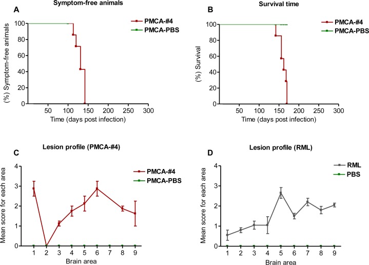
Fig 9. Incubation time, survival time and lesion profile analysis of PMCA-#4 infected animals.
The animals injected with PMCA-#4 showed an incubation (A) and survival time (B) of 130 ± 4.4 and 160 ± 3.85 days (Mean ± Standard Error of the Mean, SEM) and results were analyzed with the Logrank test. Spongiform profiles were determined on Hematoxylin and Eosin (H&E)-stained sections, by scoring the vacuolar changes in nine standard gray matter areas: 1. Dorsal medulla; 2. Cerebellar cortex; 3. Superior culliculus; 4. Hypothalamus; 5. Thalamus; 6. Hippocampus; 7. Septum; 8. Retrosplenial and adjacent motor cortex; 9. Cingulated and adjacent motor cortex, as described by Fraser et al. [54]. Lesion profile was compared to that of RML (i.c.) infected animals. Bars in C and D indicate Standard Error of the Mean.

