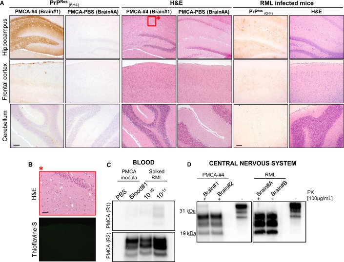
Fig 10. Detection of PMCA-#4 in brain and blood of infected animals and related immunohistochemical/biochemical characterization.
Neuropathological analysis of PMCA-#4 and PMCA-PBS injected animals and comparison to that of RML infected mice. Animals injected with PMCA-#4 showed widespread deposition of PrPRes in the hippocampus with focal plaque-like deposits found in the cerebral cortex. Cerebellum is completely spared by PrPRes accumulation. RML injected animals showed the typical pattern of widespread, synaptic and diffuse PrPRes accumulation in the whole brain with major involvement of thalamus and hippocampus. Spongiform changes were mainly found in the hippocampus of PMCA-#4 injected animals. Few vacuoles were detected in the cerebral cortex, while cerebellum did not show any vacuolation. RML injected animals showed severe vacuolation in the thalamus and hippocampus. Mild alterations were found in cerebellum and septum. Brain of PMCA-PBS injected animal was used as control (A). Higher magnification of the red square in panel A (see asterisk) and Thioflavin-S staining showing the lack of amyloid properties of the deposits found in the submeningeal level of the cerebral cortex (B). PMCA of blood collected at 140 day post infection (d.p.i.) from a symptomatic animal injected with PMCA-#4. RML dilutions (10−10 and 10−11) were used in PMCA to estimate the concentration of circulating infectious PrP (C). Biochemical analysis of the brains harvested from the first two animals injected with PMCA-#4 (sacrificed at terminal stage of the disease) were performed and compared to that of RML injected mice (D). Scale bar in A is 10 μm; scale bar in B is 5 μm. Western blots were performed using 6D11 monoclonal antibody to PrP (0.2 μg/mL, Covance). Blots were developed with the enhanced chemiluminescent system (ECL, Amersham Biosciences) and visualized using a G:BOX Chemi Syngene system.

