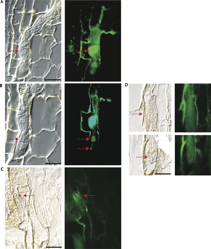
Fig 6. Immunolocalization of HgSNARE-like protein-1 (HgSLP-1).
Panels A—D are 40 x light field images matched with corresponding epiflorescent images of sections of SCN in soybean roots stained using HgSLP-1 antibodies. Arrows point to the basal cell of a subventral esophageal gland in A, the median bulb and esophageal lumen in B and the stylet in C. Panel D shows negative control sections lacking HgSLP-1 antibody staining in the nematode. Arrows in D point to the basal cell of an esophageal gland and the stylet. Panel D is a composite of two sequential sections from the same nematode. For all light field images, 20 micron scale bars are shown.

