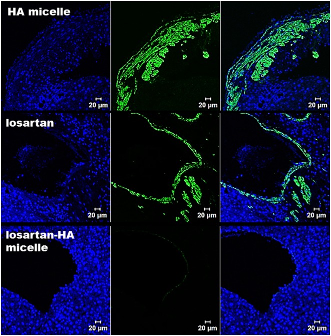
Fig 7. Confocal microscopy imaging of α-sma expression in liver section.
In HA-micelle group, mice injected with HA micelle shows high expression of α-sma as the fibrotic state of liver have increased proliferation of activated HSC which express α-sma (represented by green fluorescence). In losartan group mice injected with losartan reveals similar expression profile of α-sma compared to HA micelle treated mice group as the oral losartan have failed to reach the target region (activated HSC). In losartan-HA micelle group, mice injected with losartan-HA micelle have almost no expression of α-sma which indicate successful delivery of losartan encapsulated losartan-HA micelle in the target region. Blue color indicate DAPI stain.

