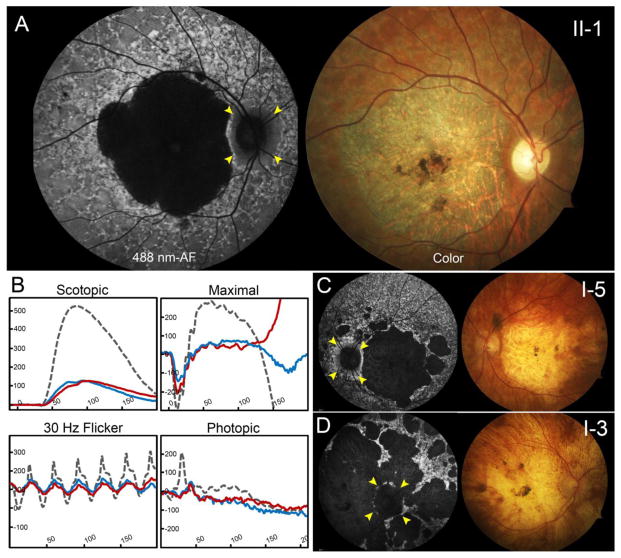
Figure 2.
Advanced ABCA4 phenotype of the proband, paternal aunt and uncles. (A) The proband, a 43-year-old man of German descent harboring the p.C54Y and c.[302+68C>T;4539+2028C>T] variants, presented with large well-delineated lesions of chorioretinal atrophy and central pigment clumping in both eyes on autofluorescence imaging and color fundus photographs. Surrounding areas of dense hyperautofluorescence preceding a reticular pattern of granular flecks were found extended throughout the periphery. (B) Full-field electroretinogram testing in the proband (II-2) revealed significantly reduced amplitudes in rod and cone responses in the right (blue trace) and left (red trace) eyes when compared to an age-matched control (dotted gray trace). The paternal aunt (c.[302+68C>T;4539+2028C>T]; c.6148-698_c.6670del/insTGTGCACCTCCCTAG) presented with similarly large area of chorioretinal atrophy while the paternal uncle (D) with the same compound heterozygous genotype exhibited generalized atrophy of the posterior pole. Various degrees of sparing of the peripapillary region in each patient are marked with yellow arrowheads.

