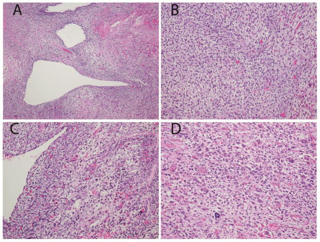
Fig. 1.
Pathologic features of the primary ovarian sarcoma in Patient #1 with DICER1 mutation
a. Low power view shows epithelial lined cysts, solid sarcoma and focal necrosis. b. Higher power view shows spindled and stellate cells in a pale mucoid matrix. c,d. Higher power views highlight areas with significant nuclear pleomorphism. (Hematoxylin and eosin; x100 (a), x200 (b,c,d)).

