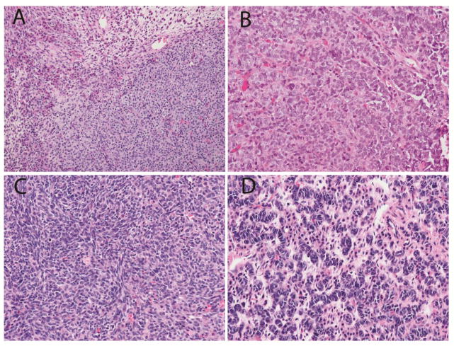
Fig. 2.
Sertoli-Leydig cell tumor in the contralateral ovary of Patient #1.
a. Low power view showing Leydig cells with abundant pink cytoplasm in upper left adjacent to nests of small Sertoli cells. b. Elongated tubules of Sertoli cells. c. Spindle cell pattern seen focally. d. Ribbons of Sertoli cells with hyperchromatic nuclei. (Hematoxylin and eosin; x100 (a), x400 (b,c,d))

