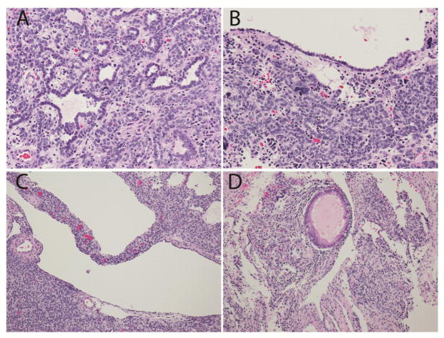
Fig. 3. Pathologic features of the Sertoli-Leydig tumor in patient #2.
a.Tubular structures and nests of Sertoli cells. b. Epithelial lined cyst with nest of Sertoli cells and rare anaplastic cells. c. Cystic structure within the SLCT. d. Focal mucinous gland. (Hematoxylin and eosin; x200 (a,b), x100 (c,d)).

