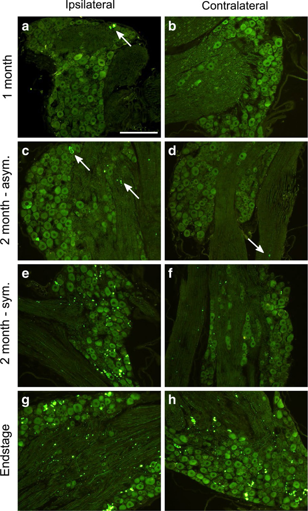
Fig. 3.
Accumulation of SOD1:YFP pathology over time in the DRG of sciatic nerve injected animals. The L3, L4, and L5 DRG were dissected from mice at various timepoints p.i. (n = 3–4 per timepoint) from both the ipsilateral (left column) and contralateral (right column) sides of the spinal column, and processed for paraffin embedding. Following deparaffinization, direct YFP fluorescence was imaged. G85R-SOD1:YFP inclusion pathology was first observed at 1 month p.i. (a, b) in the ipsilateral DRG (indicated by arrow) and grew more abundant in both the asymptomatic (c, d) and symptomatic (e, f) mice at 2 months p.i. g, h The mice at the end-stage of disease had abundant pathology in their DRG both ipsilateral and contralateral to the side of inoculation. Inclusions indicated by arrows. Scale bars 200 µm

