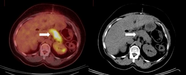
Figure 2. Postoperative initial PET/CT scan.

There is an FDG-avid lesion in the pancreatic tail on the PET axial image (white arrow, left image) with a corresponding hypodense correlate on the non-contrast enhanced CT axial image acquired as part of the same study (white arrow, right image).
