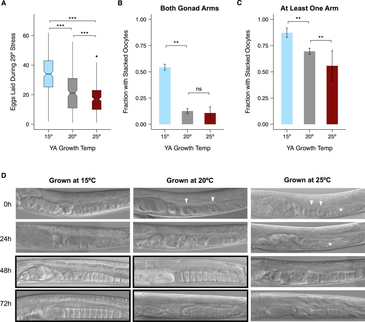
Fig 6. Maternal growth temperature affects damage sustained by the gonad during 29°C heat stress.
(A) Number of eggs laid during a 24-hour exposure to 29°C by hermaphrodites raised at different temperatures. Only those worms that laid during the stress are represented. Black lines represent median values. Box hinges represent the first and third quartiles of the data. Whiskers extend to 1.5 × inter-quartile range. (B) Fraction of hermaphrodites with stacked oocytes in both gonad arms after 48 hours of recovery from stress at 29°C. (C) Fraction with stacked oocytes in at least one gonad arm after 48 hours of recovery. (D) Representative images of worms raised at different temperatures during recovery. Images displaying stacked oocytes are boxed in black. White arrowheads point to rounded oocytes and white asterisks highlight disorganized regions of the proximal gonad. Significance levels in (A) were calculated with the Mann-Whitney U-test and in (B, C) with the binomial exact test, both with the Bonferroni-Holm correction for multiple testing (**p < 0.01, *** p < 0.001, ns: p > 0.05). Error bars represent ±1 SEM. See S7 and S9 Figs for individual trials and S3 Table for raw data.

