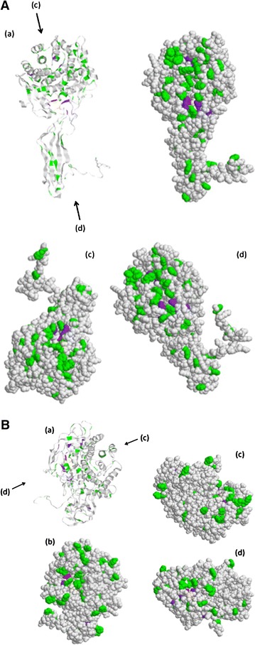
Fig. 4.

The difference of cleft shapes between EngH (A) and EngK (B). Three-dimensional models for EngH and EngK based on homologues of known structure were built by 3D-JIGSAW. The models were visualized using Rasmol tool. The aromatic amino acids, histidine and putative catalytic base in the models of the ribbon diagram (a) and the space-filling (b, c, d) were colored green, purple and red, respectively. The arrows indicate the direction of looking in the cleft
