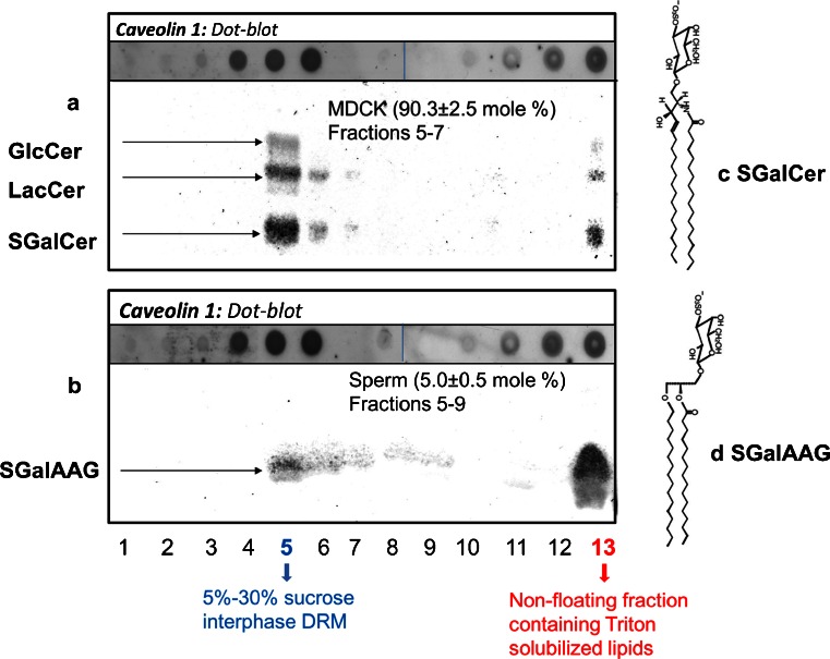
Fig. 2.
Partitioning of glycolipids and caveolin-1 in the sucrose gradient of 1 % Triton X-100 at 4 °C treated MDCK cells and sperm. The sucrose gradient of MDCK cells and sperm (cf. Fig. 1) was divided into 13 fractions of 1 ml. Proteins of fractions 1–13 were solubilized and transferred to a PVDF membrane (dot blot). Specific antibody binding was detected with enhanced chemifluorescence. For presentation purposes, dots of fractions 9–13 were aligned aside the spots of 1–8; the dots were originally spotted in multiple rows of 8 dots on one PVDF membrane and developed in the same fashion. Lipids from the 1–13 fractions were extracted, from which the glycolipids were purified and spotted on HPTLC plates, which was after development and charred with orcinol to allow purple staining of glycolipids (for method, see Gadella et al. 1993). a Dotblot and HPTLC for MDCK cells and b for boar sperm cells. The amount of sulfatides (SGalCer for structure: c for MDCK and seminolipid; SGalAAG for structure: d of fraction 13 versus fraction 5–9) was quantified according to the coloric method of Kean (1968) as modified by Radin (1984). Mean values ± SD are provided (n = 5)

