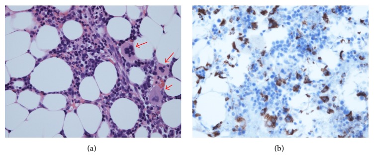
Figure 2.
(a) The phagocytic reticular cells (arrows). The phagocytosis of mature neutrophils by phagocytic reticular cells is present. Hematoxylin and eosin (HE) staining. (b) Immunohistochemistry showing monocyte infiltrations in bone marrow. The CD68+ cells increased and were diffusely distributed in stroma of bone marrow. Immunohistochemical staining using antibodies against CD68.

