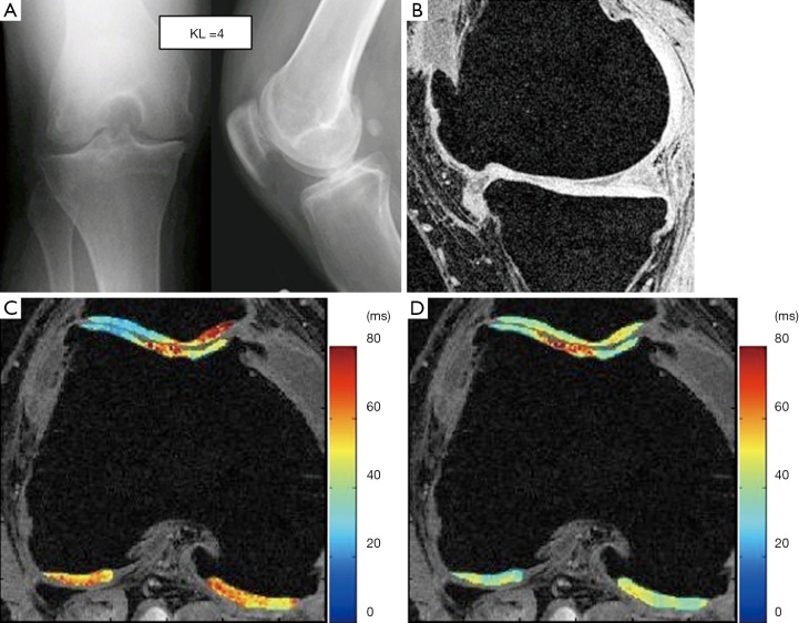
Figure 9.
Radiographs (A), T1-weighted water excitation SPGR image (B), T1ρ map (C) and T2 map (D) for a patient with advanced OA (male, 46). Based on radiographs, the patient had joint space narrowing with 1 mm in medial compartment and 3 mm in lateral compartment, and significant osteophytes in both femoro-tibial and femoro-patellar joints, resulting in a KL score as 4. In MR images, significant osteophytes were seen in both femoro-tibial and femoro-patellar joints. The cartilage had a grade 3 thinning in medial femur, medial tibia and femoro-patellar compartments, and grade 2 thinning in lateral femur and lateral tibia compartments. The average T1ρ value was 55.4±26.0 ms and the average T2 value was 43.8±11.1 ms in cartilage [Reprinted with permission (78)]. OA, osteoarthritis.

