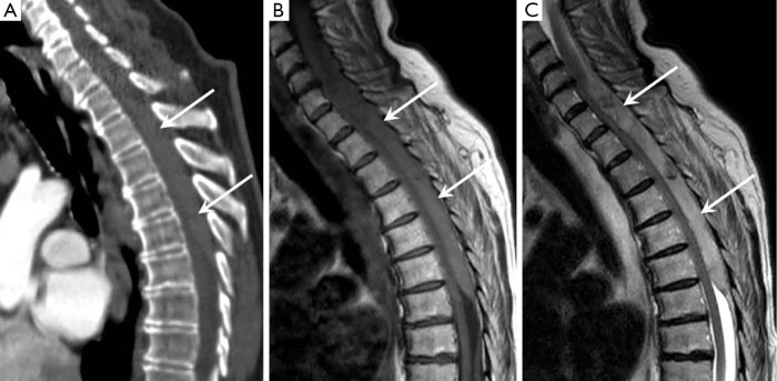
Figure 1.
(A) Sagittal CT reconstruction showing a posteriorly sited elliptical 15 cm high attenuation collection compressing the spinal cord in keeping with a large epidural haematoma (arrows); (B) and (C) sagittal T1 and sagittal T2 weighted MRI images showing the collection to be of intermediate T1 and high T2 signal intensity in keeping with a subacute haematoma. CT, computed tomography; MRI, magnetic resonance imaging.

