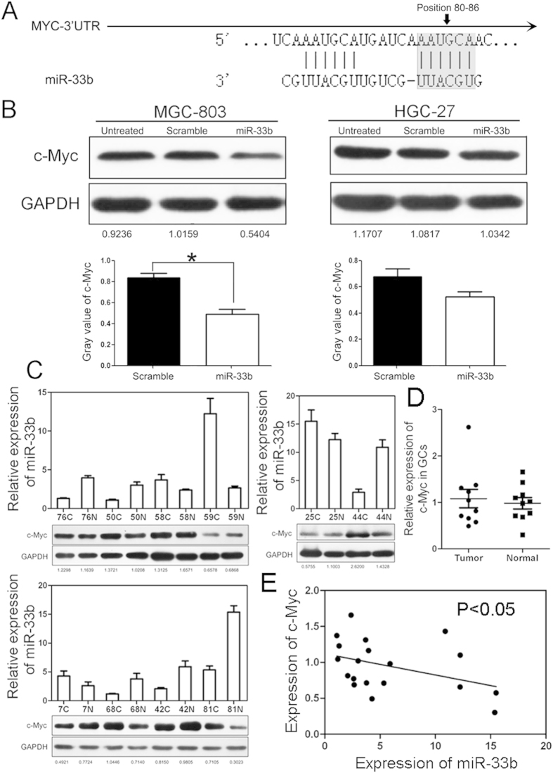
Figure 3. miR-33b targets c-Myc in GC cells and patients.
(A) Schematic representation of c-Myc 3′ UTRs showing putative miR-33b binding sites. (B) Western blot analysis of c-Myc expression in MGC-803 and HGC-27 cells transfected with scramble oligonucleotide or miR-33b mimics (Top). Gray value of c-Myc expression was also shown (Bottom). (C) Western blot analysis of c-Myc expression and relative level of miR-33b in 10 pairs of GC tissues. (D) Statistical expression of c-Myc in GC samples. (E) Expression of miR-33b was negatively correlated with c-Myc in GC sample.

