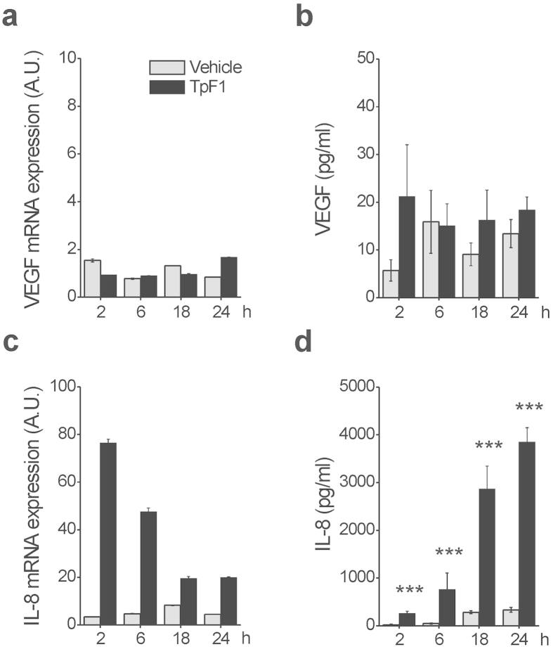
Figure 2. TpF1 stimulates IL-8 expression in endothelial cells.
HUVECs were exposed to TpF1 or vehicle (saline) for 2, 6, 18, 24 h and the expression of VEGF (a) or IL-8 (c) was evaluated by RT-PCR. Data were normalized to an endogenous reference gene (ribosomal subunit 18S). Values at T0 cells were taken as reference and set as 1 A.U. and the expression levels for treated cells were relative to the expression of T0 cells. Culture supernatants from HUVECs, harvested for quantification of mRNA, were collected and the VEGF (b) and IL-8 (d) protein content was quantified by ELISA. Data are expressed as mean ± S.D. of four independent experiments. Significance was determined by Student’s t-test for data of TpF1 treated cells versus vehicle-exposed cells. ***p < 0.001.

