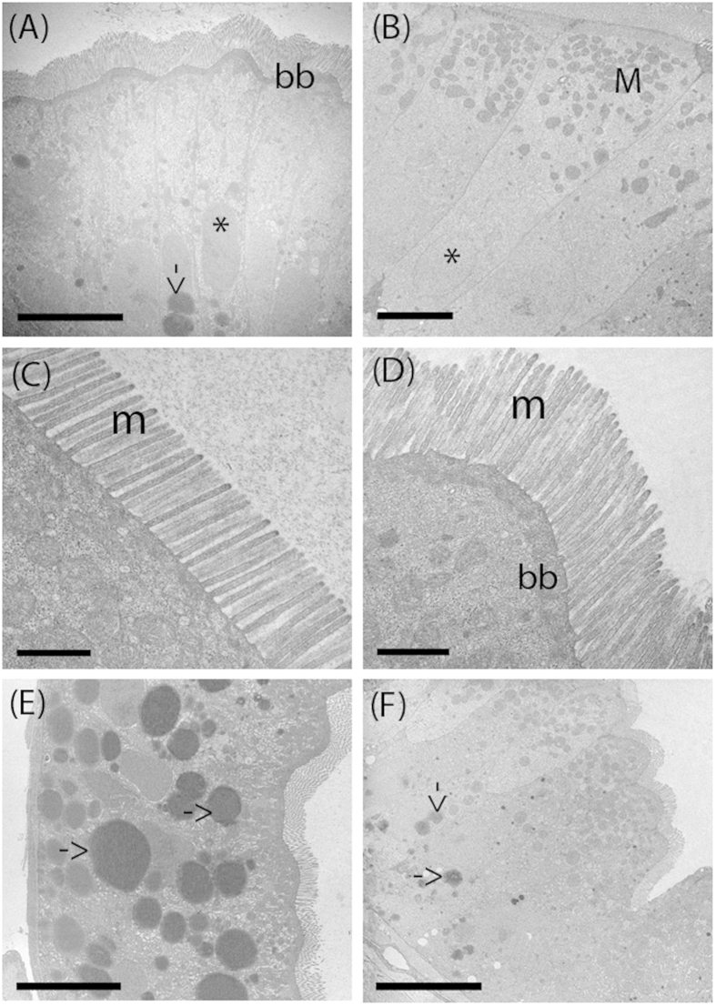
Figure 4. Transmission Electron Microscopy (TEM) shows the ultrastructure of the intestinal in control and probiotic treated zebrafish.
Thin sections of 8 dpf zebrafish showing columnar epithelia with apical brush border in control (A) and probiotic treated intestine (B). The treated intestines present abundant spherical mitochondria located in the apical part of the enterocytes, close to the brush border. Both control and treated group larvae intestine show undamaged epithelial barrier and absence of cell debris. Electron micrographs show organized microvilli on the apical surface in the enterocyte of control (C) and treated larvae (D) and the presence of lipid droplets in the enterocytes cytoplasm of control (E) and treated larvae (F). BB: brush border; L: lumen; M: mitochondria; m: microvilli, arrow: lipid droplets; *nucleus. Scale bar: 10 μm in (A); 5 μm in (B, E, F); 1 μm in (C,D).

