Figure 4.
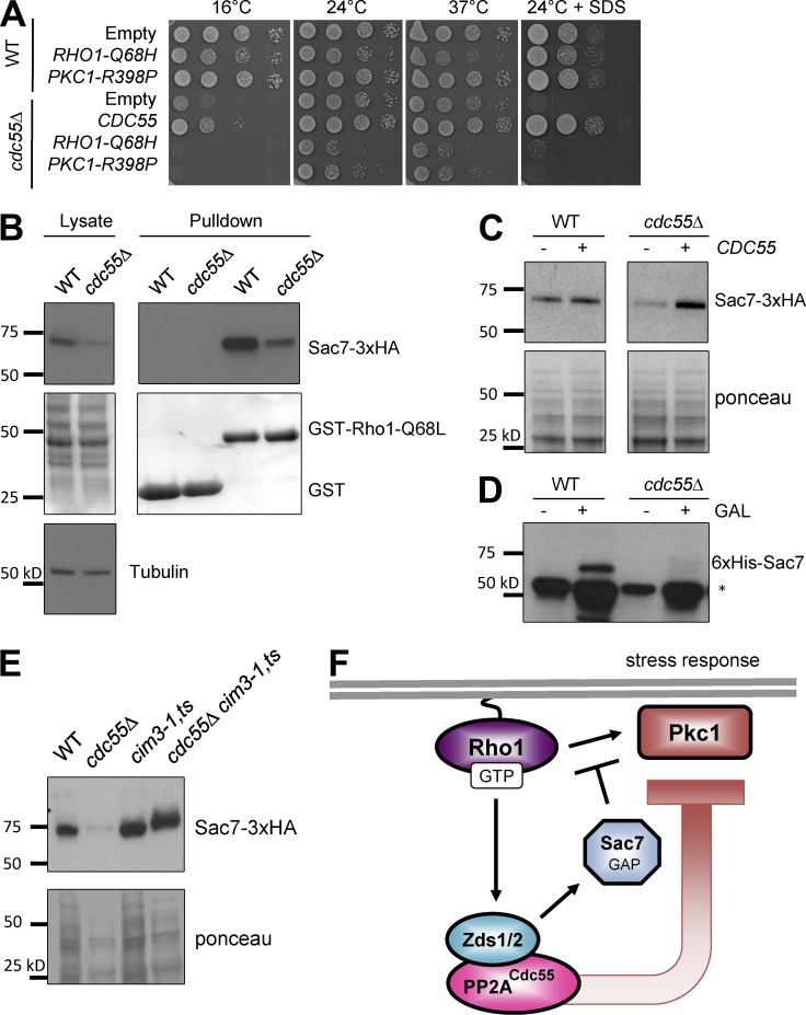
PP2ACdc55 inhibits Pkc1 activation potentially through stabilization of the Pkc1-specific Rho1 GAP, Sac7. (A) Expression of RHO1-Q68H or PKC1-R398P was toxic to cdc55Δ cells. (B) Sac7-3XHA is less abundant in the cdc55Δ lysates. Lysates and bead-bound fractions were subjected to SDS-PAGE and Western blotting with anti-HA antibody and anti–α-tubulin as a loading control. (C) Expression of CDC55 restored Sac7-3xHA protein levels in cdc55Δ cells to wild-type (WT) levels. (D) The reduction of Sac7 in the cdc55Δ lysates is a posttranscriptional mechanism. Expression of 6xHis-Sac7 from an inducible GAL1 promoter failed to accumulate Sac7 in cdc55Δ cells. *, nonspecific band detected by anti-His antibody (Millipore). (E) Sac7 is degraded by the proteasome. Inactivation of the proteasome by a cim3-1 mutation at the semipermissive temperature of 30°C for 4 h resulted in stabilization of Sac7-3xHA even in the cdc55Δ mutants. (F) Model for how Zds1/Zds2–PP2ACdc55 antagonizes Rho1 activation of Pck1 through the stabilization of the Rho1 GAP Sac7.

