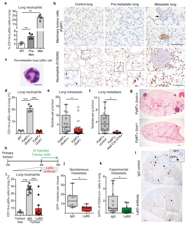
Figure 1. Neutrophils infiltrate pre-metastatic lungs and favour metastasis.
a, b, Analysis of wild-type (WT) or MMTV-PyMT+ mice. a, Lung neutrophils frequencies determined by flow cytometry (n = 5 (wild-type), n = 4 (pre-metastatic lung), n = 4 (metastatic lung)). Met., metastatic. b, Lung neutrophils or cancer cells determined by histology staining for S100A9 or PyMT (brown). Scale bars, 100 μm. Magnifications in inserts. c, Haematoxylin & eosin (H&E)-stained neutrophil. Scale bar, 5 μm. d, Lung neutrophil quantification by flow cytometry (n = 5 (wild-type), n = 4 (PyMT+ Gcsf+/+), n = 7 (PyMT+ Gcsf−/−)). e, f, Spontaneous metastasis of MMTV-PyMT+ Gcsf+/+ (n = 13) or MMTV-PyMT+ Gcsf−/− (n = 24) (e) and MMTV-PyMT+ control (n = 14) or MMTV-PyMT+Ela2-Cre-DTA+ (n = 6) mice (f). g, Representative H&E-stained sections of lung. Scale bar, 500 μm. h, Experimental setup for neutrophil depletion. i, Flow cytometric lung neutrophil quantification (n = 4 (tumour-free), n = 12 (IgG tumour), n = 11 (Ly6G tumour)). j, k, Spontaneous (n = 8 per group) (j) and experimental metastasis (n = 12 per group) (k). Lin, CD45 CD31 TER119. l, Histological GFP-stained lung sections including close-up on spontaneous (arrow) and experimental metastases (brown). Scale bar, 500μm. Statistical analysis by two-sided t-test. Data are represented as mean ± standard error of the mean (s.e.m.). *P < 0.05, **P < 0.01, ***P < 0.001.

