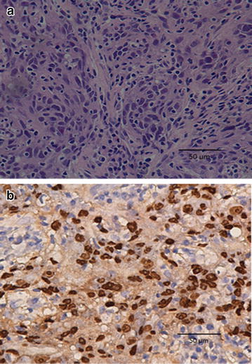
Fig. 1.

Representative images of pathologic slices from fine-needle aspiration of the occipital lymph node (original magnification, ×40). a Hematoxylin and eosin (H and E) stained section shows diffused tumor cells displaying characteristics of nasopharyngeal carcinoma (NPC) cells. b Immunohistochemical analysis and in situ hybridization of the occipital lymph node shows the expression of Epstein-Barr virus-encoded RNAs (EBERs) in tumor cells
