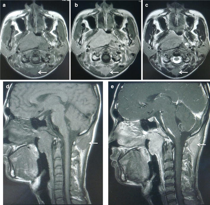
Fig. 2.

Magnetic resonance (MR) imaging of the NPC patient before treatment. T1-weighted axial MR images a without contrast, b with contrast, and c T2-weighted axial MR image show an occipital lymph node (18 mm × 19 mm) with equal T1 signal, long or equal T2 signal, and obvious enhancement (arrows). T1-weighted sagittal MR image d without contrast and e with contrast also show an enlarged lymph node with enhancement in subcutaneous tissue of the occiput (arrows)
