
Fig. 1.
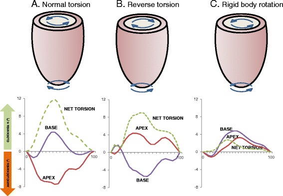
Rotational mechanics in NICM. Diagrammatic representation of torsional and rotational patterns identified using feature-tracking cardiovascular magnetic resonance. In the bottom tiles, the time in the cardiac cycle, expressed as a percentage of the R-R interval on the ECG, is shown in the x axes. Rotation at the base and apex of the LV as well as net torsion (the instantaneous difference between apical and basal rotation) is shown on the y axis (in degrees) a shows a preserved torsional pattern from a patient with non-ischemic dilated cardiomyopathy without MWF with predominantly anticlockwise rotation at the apex and clockwise rotation at the base. b shows reverse torsion, where the direction of both apical and basal rotation is reversed. c shows rigid body rotation in a patient with NICM and MWF. The apex and base both twist in the same direction so that the heart rotates as one solid body with minimal net torsion