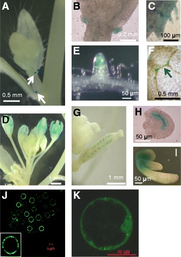
Fig. 1.

AtNUP62 promoter activity and protein distribution. Tissue-specific activity of the AtNUP62 promoter was investigated by histochemical analysis of GUS staining (blue color) in transgenic plants expressing GUS under control of AtNUP62 promoter region. a Stipules at the basis of cauline leaves (arrows). b Enlarged view of stipules at the basis of a cauline leaf. c margin of a cauline leaf. d Inflorescence. e Root tip. f Cotyledon tip. The arrows indicates the localisation of GUS staining. g Young silique and h developing seed from this silique. i Young germinating seedling. j and k, Subcellular localization of AtNUP62::GFP fusion protein (confocal microscopy). The AtNUP62::GFP construct was expressed under control of the cauliflower mosaic virus 35S promoter, and the same construct was used for plant and protoplast transformation. j Root tip of a transgenic 35S::AtNUP62-GFP 10-day old Arabidopsis plant. k Confocal microscopy analysis of AtNUP62::GFP signals in a transiently transformed Arabidopsis cultured cell protoplast
