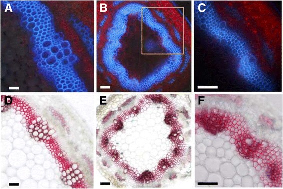
Fig. 7.

Characterization of the mtirx1-1 mutant by UV microscopy and phloroglucinol staining. a to c, stem cross sections of wild type (a), and the mtirx1 mutant (b) were observed under UV light. c A higher magnification of the rectangle marked area in (b). Collapsed xylem phenotype is evident. The blue color is the auto-fluorescence of lignin under UV light, and the red color is the auto-fluorescence of chloroplasts. d to f, cross sections of wild type (d) and the mtirx1 mutant (e) after phloroglucinol staining. f, Staining of the mtirx1 mutant at a higher magnification. Bars = 10 μm
