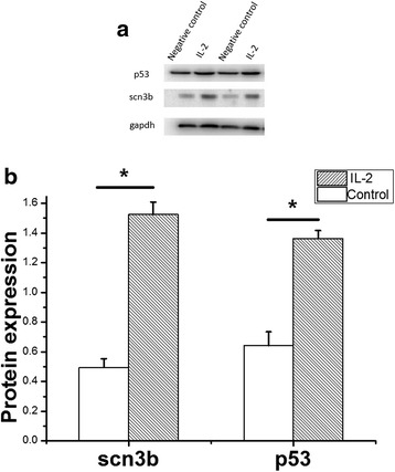
Fig. 5.

The expression of p53 and scn3b in HL-1 cells induced by Interleukin 2 (IL-2) by Western blot analysis. The protein samples were prepared from transfected HL-1 cells. Gapdh was used as a control for normalization. P53 and Scn3b were significantly increased induced by IL-2 compared with negative control. The images of Western blot analysis shown in (a) were scanned, quantified and plotted in (b). Data is shown as means and SD
