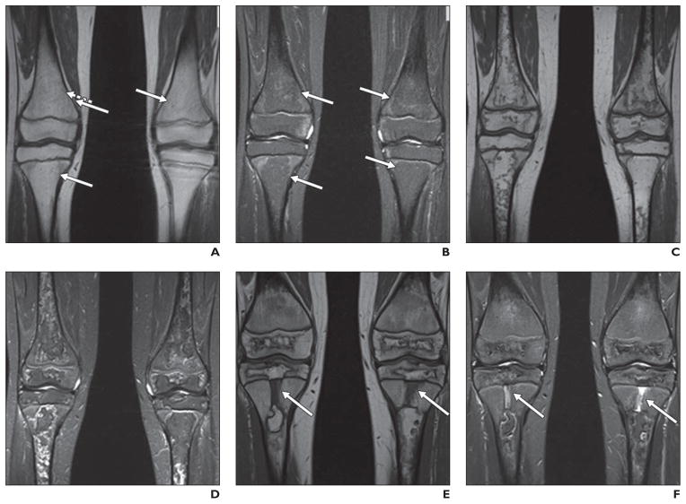
Fig. 1. 9-year-old girl being treated for standard-risk acute lymphoblastic leukemia who underwent routine MR screening for osteonecrosis.
A and B, Coronal unenhanced T1-weighted (A) and STIR (B) images of knees show very subtle geographic signal changes (arrows) in distal femoral and proximal tibial diametaphyses bilaterally. These signal changes are less well defined than those described in literature for osteonecrosis.
C and D, MR images show that over course of 3 months, these changes evolved into typical geographic signal changes of osteonecrosis.
E and F, Annual follow-up examinations (not shown) revealed progressive definition of these extensive lesions, with overall decrease in extent. Note rectangular foci of abnormal centrally located metaphyseal signal in proximal tibias (arrows), dark on T1-weighted image (E) and bright on STIR image (F). Over course of 3 years, these abnormalities narrowed transversely and elongated, coincident with longitudinal growth of patient. These findings represent cartilaginous ingrowths from arrested enchondral ossification.

