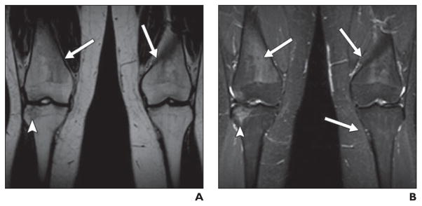
Fig. 4. Mottled marrow pattern in 18-year-old man previously treated for acute lymphoblastic leukemia who underwent MRI for complaint of chronic knee pain.
A and B, Coronal unenhanced T1-weighted (A) and STIR (B) images of knees show bilateral heterogeneous marrow signal (arrows) in distal femoral and proximal tibial metaphyses, which is mildly dark on T1-weighted and bright on STIR images. These changes became less apparent over course of 2 years (not shown). Also note area of nonspecific edema (arrowheads) in right lateral tibial epiphysis, which completely resolved over 2 years.

