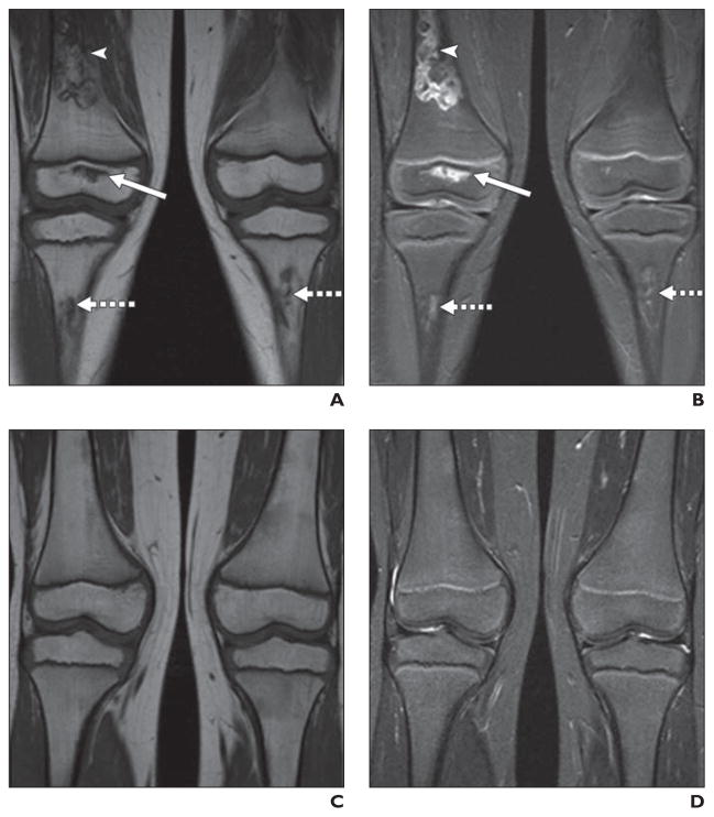
Fig. 7. Healing osteonecrosis of knees in 4-year-old girl undergoing treatment of acute lymphoblastic leukemia who was prospectively monitored annually for osteonecrosis of hips (normal throughout monitoring) and knees.
A and B, Coronal unenhanced T1-weighted (A) and STIR (B) images of knees show small lesion of right distal femoral epiphysis (solid arrows), large lesion of distal right femoral diaphysis (arrowheads), and moderate-sized lesions of proximal tibial diametaphyses bilaterally (dotted arrows).
C and D, Over course of 4 years, osteonecrotic lesions healed. Coronal unenhanced T1-weighted (C) and STIR (D) images of knees show no evidence of osteonecrosis.

