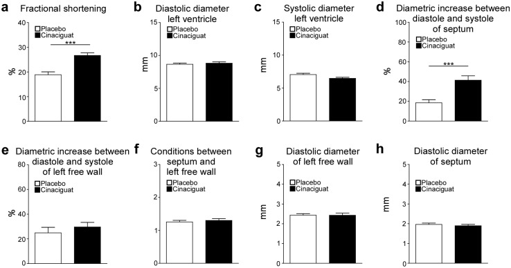
Fig 5. Results of M-Mode echocardiographic analysis performed one week before study end.
Fractional shortening (a), diastolic (b) and systolic (c) diameter of the left ventricle, diametric increase between diastole and systole of the septum (d) and left free wall (e), conditions between septum and left free wall (f) as well as diastolic diameter of left free wall (g) and septum (h) were analysed in 14 (placebo) and 16 (cinaciguat) animals. Data are means ± SEM. ***p < 0.005: Student’s t-test (cinaciguat vs. placebo). 14 animals of the placebo group and 16 animals of the cinaciguat group were analyzed.

