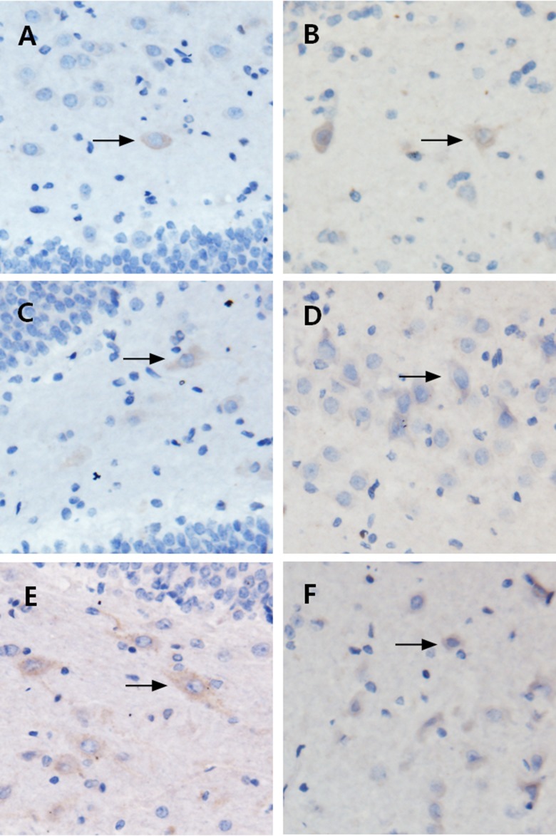
Figure 2.
Localization of ANXA1 via immunohistochemistry. A (control), B (24 h), C (72 h), D (1 week), E (2 weeks), and F (1 month). Immunohistochemically, the glial cells in the hippocampus stained positive for ANXA1, as demonstrated by brownish yellow-stained particles distributed in the cytoplasm. (A) The control group, showed brownish yellow particles with weakly positive staining; (B–F) The model groups showed brownish yellow particles with deep staining, especially in B and F. The ANXA1 expression was more pronounced in the model groups compared to the control group.

