
Fig. 2.
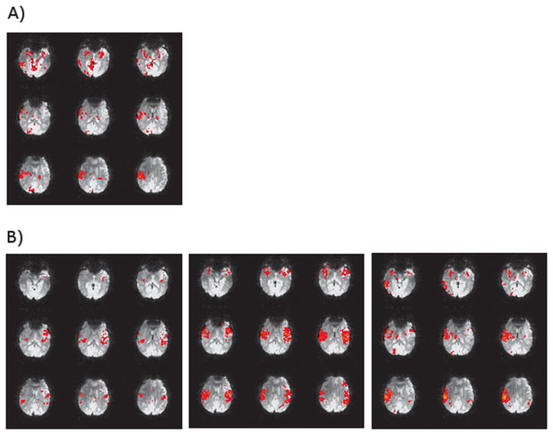
EEG/fMRI analysis implemented in FSL’s FEAT with standard preprocessing steps. Subject was diagnosed with BECTS and had 56 spikes during a resting EEG/fMRI acquisition (26 Right, 23 Left, 7 Bilateral). Images show positive BOLD activation for right spikes with spike times convolved with A) FSL’s standard Gamma HRF (6 second lag, 3 second standard deviation) and B) a FIR basis set starting at the time of the spike with a two second window (i.e. each image is 2 seconds apart showing the progression from time of the spike to 6 seconds after).