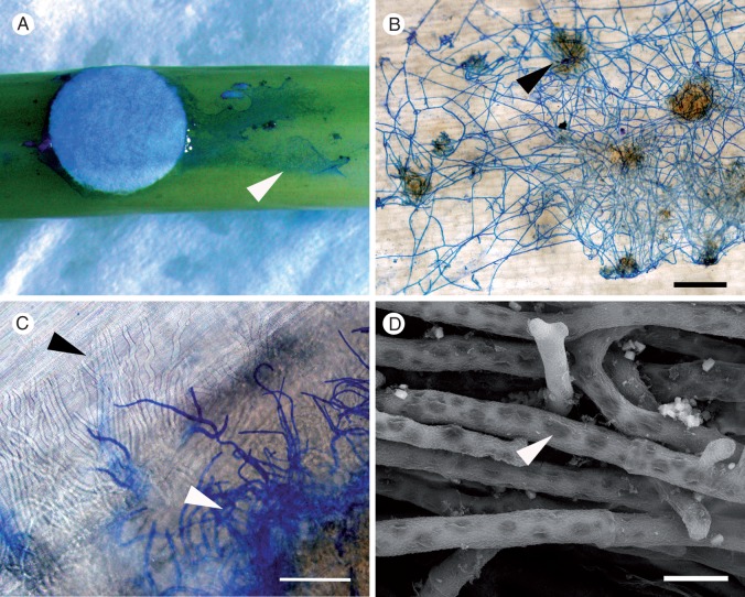
Fig. 2.
Light and scanning electron micrographs showing disease progression caused by Sclerotinia sclerotiorum MBRS-1 (A–C) and WW-3 (D) on the stems of Brassica carinata following inoculation using colonized filter paper discs. (A) Extension of S. sclerotiorum along the surface of the stem of susceptible B. carinata SMP3-82 (arrowhead) at 24 hpi. (B) Established hyphal net on the surface of resistant B. carinata 054113 showing infection cushions (arrowhead) at 48 hpi. (C) Subcuticular hyphae visible in susceptible B. carinata SMP3-82 (black arrowhead) and surface hyphae (stained blue, white arrowhead) at 48 hpi. (D) Hyphae in the lesion and on the stem surface became highly vacuolated (arrowhead) by 72 hpi (susceptible B. carinata SMP3-82). Scale bars (B) = 500 µm, (C) = 66 µm, (D) = 10 µm.

