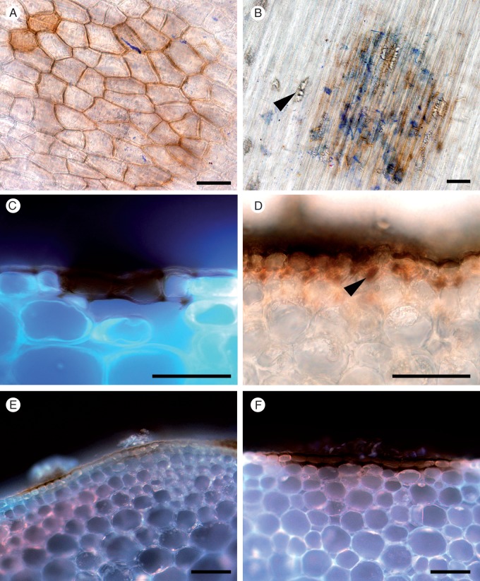
Fig. 9.
Discoloration of epidermal cell walls after resistant and susceptible Brassica carinata and B. napus lines were challenged with S. sclerotiorum isolates MBRS-1 and WW-3 (A, B, C, E, F). Hypersensitive reaction (HR) at sites of failed infection on Brassica stems. (A) Resistant B. carinata 054113 at 3 dpi (WW-3). (B) Disintegrating hyphae, calcium oxalate crystals (arrowhead) and discoloration of cell walls at the site of an attempted infection on the stem of resistant B. napus ZY006 at 3 dpi (MBRS-1). (C) Discoloration of cell walls and cell death at site of a failed infection on resistant B. carinata 054113 at 3 dpi (WW-3). (D) Successful infection, with growth of subcuticular hyphae (arrowhead) in susceptible B. napus YM04 at 3 dpi in (MBRS-1). (E, F) Discoloration of cell walls and cell death at site of a failed infection on (E) susceptible B. carinata SMP3-82 at 3 dpi (MBRS-1) and (F) resistant B. carinata 054113 at 3 dpi (MBRS-1). Scale bars (A–F) = 50 µm.

