
Fig. 1. Antigenic characteristics of the 92UG037.8 Env trimer presented on cell surfaces.
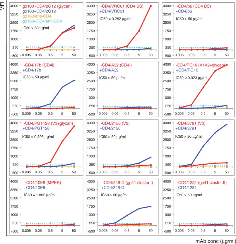
Plots of antibody binding to the Env trimer on the 92UG037.8 gp160 cell surfaces in the absence (red) or presence (blue) of soluble CD4. Fluorescent signal for bound CD4 is shown in the presence of CD4 (cyan) or in the absence of CD4 (orange). Antibodies and their epitopes are indicated. The median inhibitory concentration (IC50) values were determined in a luciferase-based virus neutralization assay using 92UG037.8 gp160 and purified antibodies. Unless specified, all antibodies used are Fab fragments. Original flow cytometry histograms are shown in fig. S4. Extensive control experiments were carried out to ensure the binding specificity, and the experiments were repeated at least twice with almost identical results.