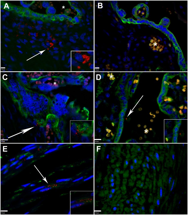
Fig 3. In situ detection of Pg in placental and umbilical cord sections from preterm and term tissues.
A. Placental villous section from a preterm specimen demonstrating the distribution of Pg (red, white arrow) within the extracellular matrix. Cytotrophoblasts/syncytiotrophoblasts (green) were detected with cytokeratin-7 specific antibody. B. Corresponding preterm placental section stained with pre-adsorbed Pg-specific antiserum (red) and cytokeratin-7 specific antibody (green). C. Pre-term placental specimen demonstrating Pg (red, white arrow) attached to cytotrophoblasts/syncytiotrophoblasts (green). D. Term placental villus section demonstrating Pg (red, white arrow) in association with syncytiotrophoblast (green). E. Preterm umbilical cord section demonstrating extracellular and internalized Pg (white arrow) within the perivascular stroma, myofibroblast (green) were labelled with vimentin specific antibody. F. Representative umbilical cord section from a term specimen, which were negative for Pg. Cell nuclei (blue) were stained with DAPI. *Indicates autofluorescent red blood cells in maternal or fetal circulation. In all panels, magnified inserts are Pg positive regions demarcated by arrows. All scale bars (bottom left corner) are equivalent to 10 μm, images are 400x or 600x magnification.

