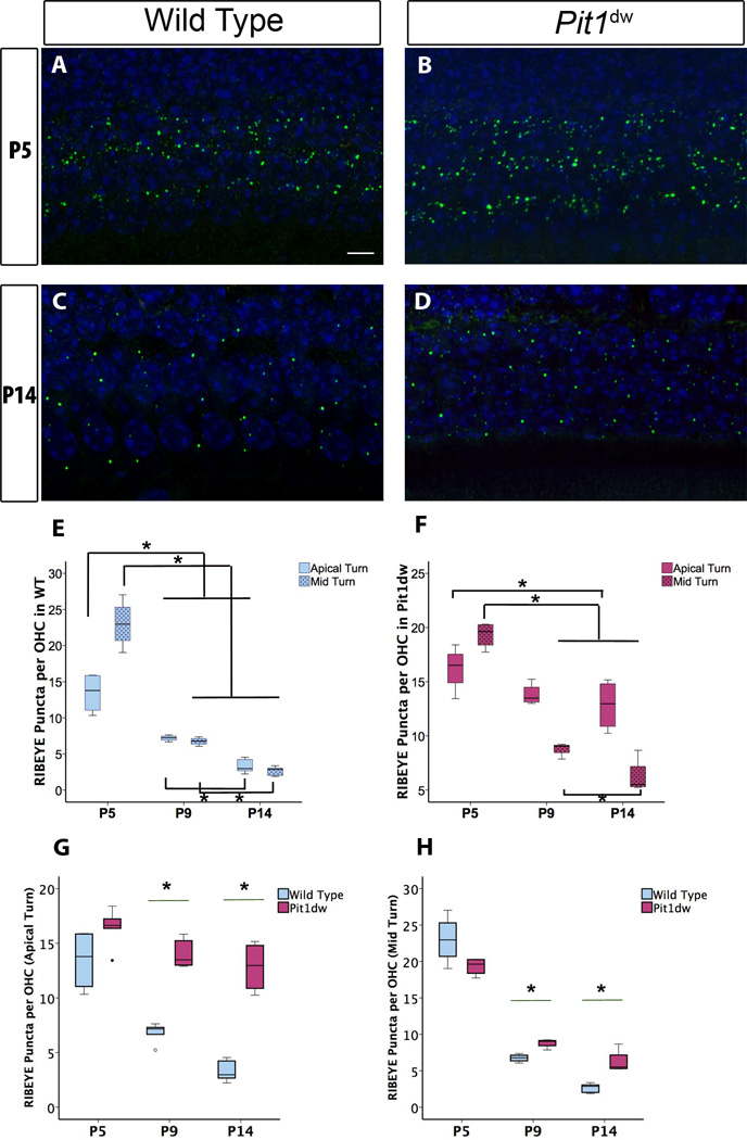
Figure 1. Outer hair cell (OHC) synaptic refinement is disrupted in Pit1dwmutants.
Projection of confocal sections obtained from the apical turn of cochlear whole mounts stained with the afferent presynaptic marker RIBEYE (in green) at P5 in wild type (WT) (A) and Pit1dw mice (B). The same marker at P14 in WT and Pit1dw mice is shown in (C) and (D) respectively. E, F, Quantification of RIBEYE puncta from the cochlea in WT and Pit1dw mice, respectively at P5, P9, and P14. G, H, Comparison of RIBEYE puncta counts in the OHCs of Pit1dw mutants at P5, P9, and P14 as compared to age-matched WT controls. Box plots represent the entire data set. Statistical tests were performed using ANOVA followed by Scheffe’s post hoc test. n≥4 for all groups. *p<0.05 was considered statistically significant. Statistically significant comparisons are indicated (*).

