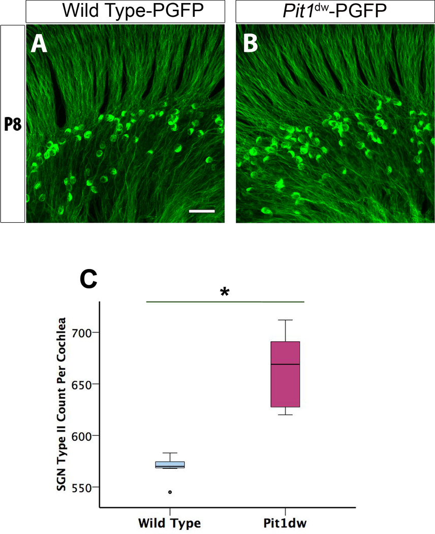
Figure 2. Altered pruning of spiral ganglion type II neurons in P8 hypothyroid organ of Corti.
A, B, Projection of confocal sections obtained from whole mount cochlea of PGFP that has type II SGNs labeled with GFP (green) (A) and PGFP-Pit1dw mice (B). C, Quantification of the SG II neurons in the organ of Corti of Pit1dw mutants compared to WT controls. Results are expressed as a box plot showing the entire data set. The scale bar represents 10 µm. n=7 for both PGFP and PGFP- Pit1dw mice. *p<0.001 by Student’s t test.

