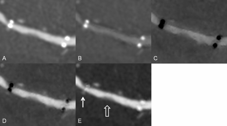
Fig 4. An intracranial stent in a patient.
Multiplanar reconstruction of (A) monochromatic 70-keV image, (B) monochromatic 140-keV, (C) iodine (calcium), (D) iodine (HAP), and (E) iodine (water) images. The radiopacity of the stent marker was reduced in the iodine (water) images (white solid arrow). A soft plaque or intimal hyperplasia is visible in the in-stent lumen (white open arrow).

