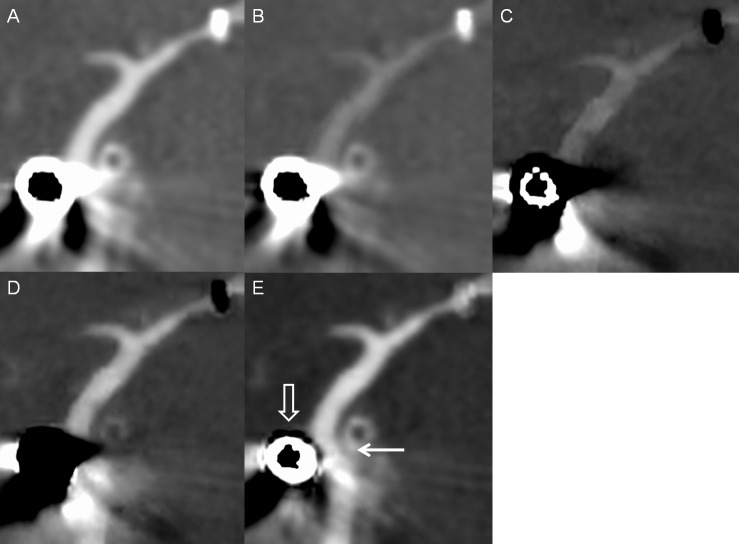
Fig 6. Intracranial stent and aneurysm coiling in a patient.
Multiplanar reconstruction of (A) monochromatic 70-keV, (B) monochromatic 140-keV, (C) iodine (calcium), (D) iodine (HAP), and (E) iodine (water) images. The vessel lumen near the aneurysm coiling (white open arrow) is visible on the iodine (water) image (white solid arrow) and invisible on other images because of severe artifacts.

