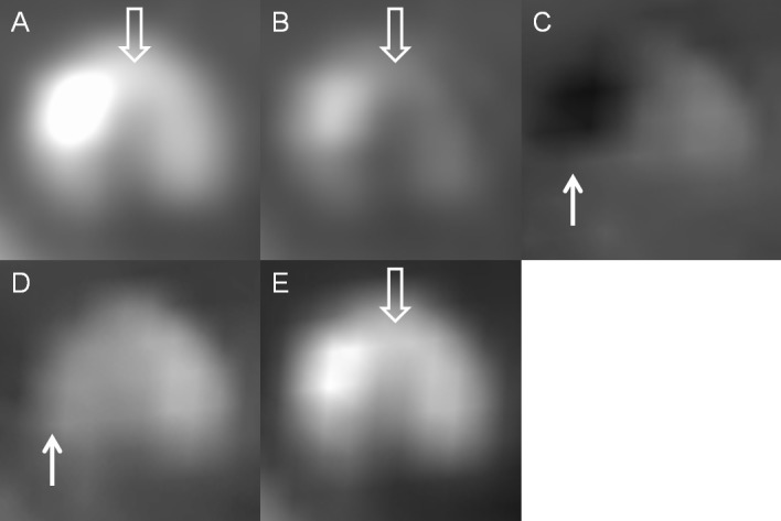
Fig 7. A mixed plaque in a patient with an intracranial stent.

Axial view of (A) monochromatic 70-keV, (B) monochromatic 140-keV, (C) iodine (calcium), (D) iodine (HAP), and (E) iodine (water) images. The calcification of mixed plaque had the appearance similar to that of the stent marker and showed dark on iodine (calcium) images and disappeared on iodine (HAP) images (white solid arrow). Iodine (water) and 140-keV images reduced the blooming artifacts caused by the calcification of mixed plaque rather than change its hyperdensity. The noncalcified portion of mixed plaque remained hypodense on each mode and was visible on 70-keV, 140-keV and iodine (water) images (white open arrow).
