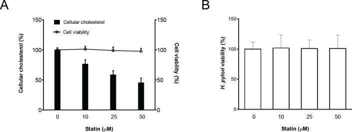
Fig 1. The level of cellular cholesterol in gastric epithelial cells was reduced through treatment with statins.
AGS cells were treated with various concentrations of simvastatin (0–50 μM) and infected with H. pylori at an MOI of 100 for 6 h. (A) Whole cell lysates were then prepared for cholesterol level analysis (open bar). (B) Bacterial suspension was plated onto Brucella blood agar plates and incubated for 3–4 days, after which the CFUs were counted for evaluation of bacterial viability (open circle). (B) Cell viability was not influenced by treatment with simvastatin, as determined by the trypan blue exclusion assay. The data are presented as means ± standard deviations for three independent experiments. Statistical significance was evaluated using Student’s t-test (*, P < 0.05).

