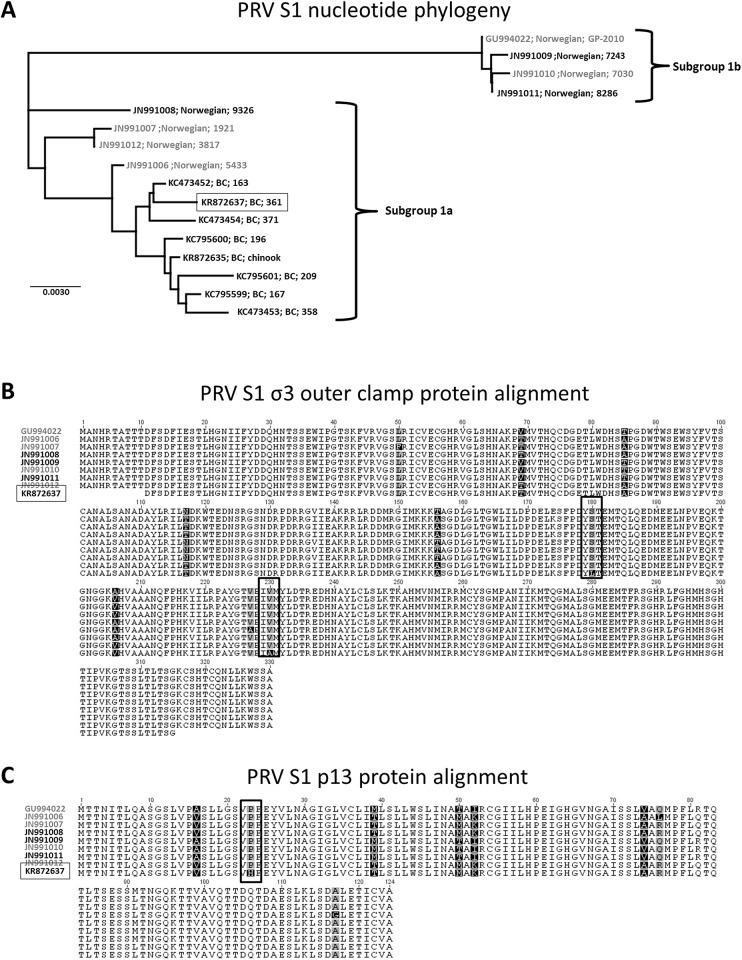
Fig 5. PRV S1 segment phylogenetic and protein comparisons between HSMI and non-HSMI associated isolates.
Phylogenetic Jukes-cantor neighbor-joining comparison of previously published sequences from Norway and Canada with the Canadian sequence (boxed) of PRV used in this study (A). Predicted amino acid alignments of both the σ3 (B) and p13 (C) proteins identify unique substitutions (vertical boxes) for the Canadian sequence of PRV in this study compared to eight Norwegian PRV sequences. In all cases, sequences of PRV obtained from HSMI disease fish (gray) are distinguished from non-HSMI associated variants (black).

