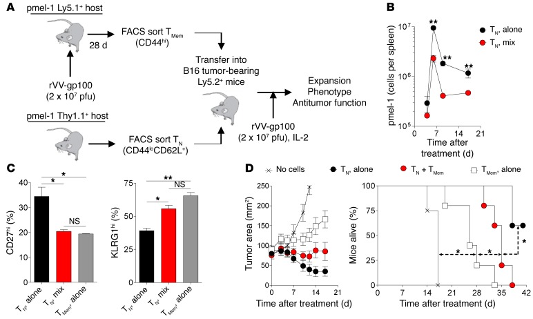
Figure 3. TMem cells cause precocious differentiation of TN cells in vivo.
(A) Experimental schema showing the generation, isolation, and transfer of Thy1.1+ pmel-1 TN cells (CD44loCD62L+) alone or in combination with FACS-sorted, vaccine-induced Ly5.1+ pmel-1 TMem cells (CD44hi) into Ly5.2+ hosts. (B) In vivo expansion and persistence of 1 × 10⁵ adoptively transferred Thy1.1+ TN cells injected alone or in combination with 3 × 10⁵ Ly5.1+ TMem cells into Ly5.2+ WT mice bearing 10-day established B16 melanomas. (C) FACS analysis on day 17 of CD27 and KLRG1 expression on CD8+Thy1.1+ TN-derived or CD8+Ly5.1+ TMem-derived cells. (D) Tumor regression and survival of mice bearing 10-day established B16 melanoma tumors who received 1 × 10⁵ TN cells alone, in combination with 3 × 10⁵ TMem cells, or 3 × 10⁵ TMem cells alone. All treated mice received 6 Gy irradiation, i.v. rVV-gp100, and 3 days of i.p. IL-2. n = 3 mice/group/time point (B and C) or n = 5 mice per group (D). Results are displayed as mean ± SEM with statistical comparisons performed using an unpaired 2-tailed Student’s t test corrected for multiple comparisons by a Bonferroni adjustment or log-rank test for animal survival. *P < 0.05; **P < 0.01. Data shown are representative of 2 independently performed experiments.

